About ChiroWorx Family Chiropractic
Dedicated to Your Spinal Health
X-Rays for Precise Diagnosis
A Clear Picture of Your Spinal Health
Contact Us Today!
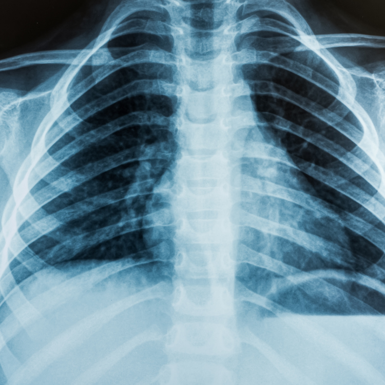
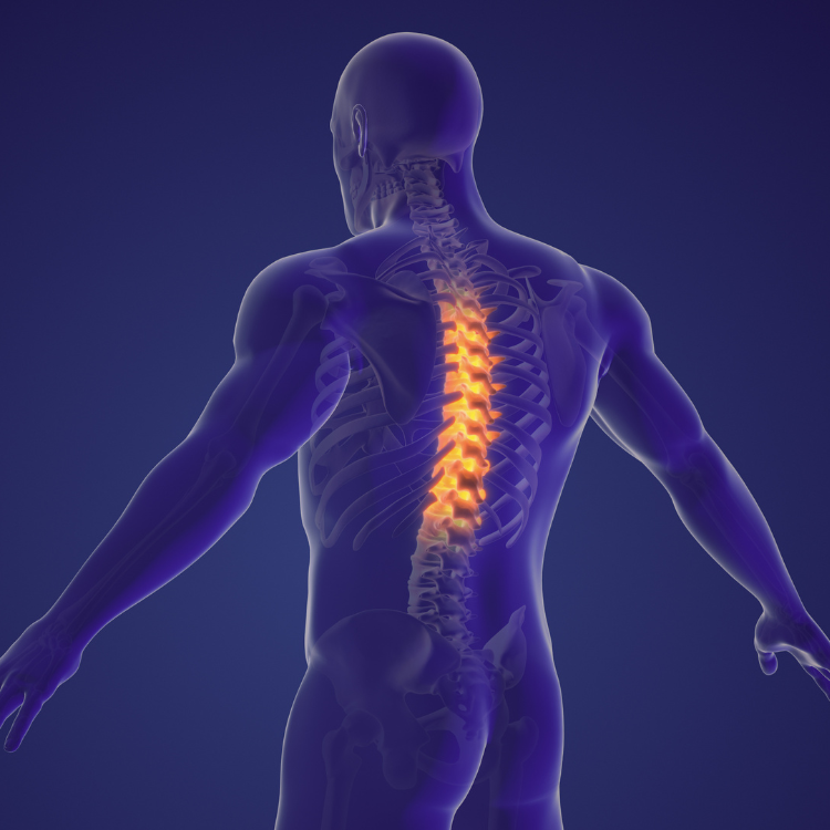
The spine is divided into four parts … So there are three common types of spinal X-Rays:
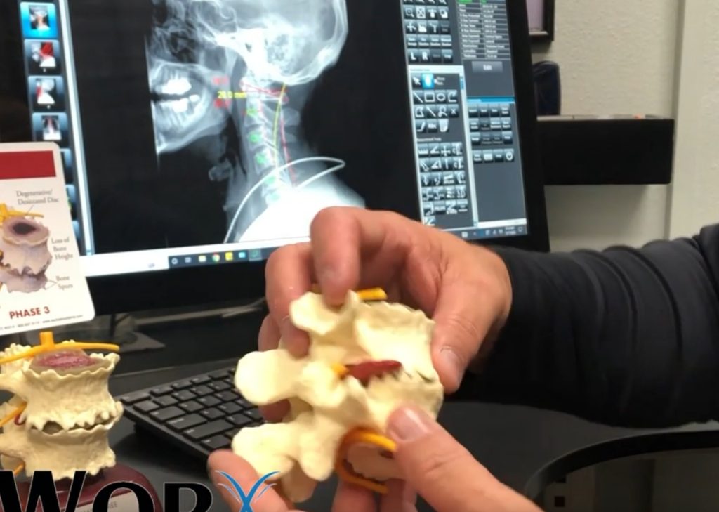
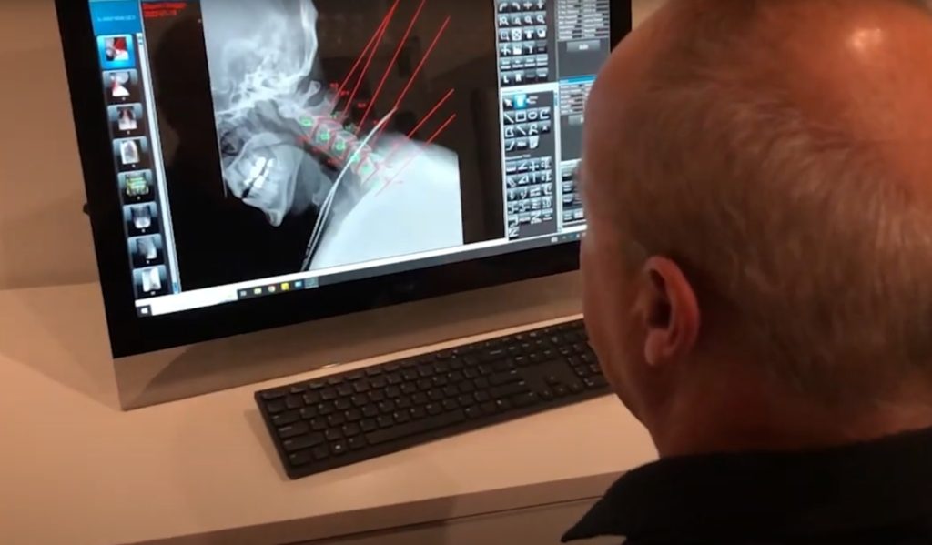
1. Cervical spine X-Ray – This X-Ray test takes pictures of the 7 neck (cervical) bones.
2. Thoracic spine X-Ray – This X-Ray test takes pictures of the 12 chest (thoracic) bones.
3. Lumbosacral spine X-Ray – This X-Ray test takes pictures of the 5 bones of the lower back (lumbar vertebrae) and a view of the 5 fused bones at the bottom of the spine (sacrum). Sacrum/coccyx X-Ray – This X-Ray test takes a detailed view of the 5 fused bones at the bottom of the spine (sacrum) and the 4 small bones of the tailbone (coccyx).
A spinal X-Ray is done to:
- Find the cause of ongoing pain, numbness, or weakness.
- Check for arthritis of the joints between the vertebrae and the breakdown (degeneration) of the discs between the spinal bones.
- Check injuries to the spine, such as fractures or dislocations.
- Check the spine for effects from other problems, such as infections, tumors, or bone spurs.
- Check for abnormal curves of the spine, such as scoliosis, in children or young adults.
- Check the spine for problems present at birth (congenital conditions), such as spina bifida.
- Check changes in the spine after spinal surgery.

